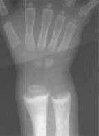
Plain film imaging (x-ray)
X-ray films or radiographs are most useful for looking at the bones and joints of the arms, legs, hands and feet. They are very useful in the chest and can be used to look at the spine in the neck, back and lower back.
Sometimes they do not give enough information and they may be used in combination with another imaging test. In some cases the radiologist may decide that a more detailed test should be used instead of plain film x-ray. For example, the investigation of headaches, and abdominal or back pain.
We try to avoid exposing pregnant patients to radiation of the abdomen or pelvis, so we may ask you if you may be pregnant.
Service: The Radiology department at Bradford Royal Infirmary serves mainly patients in hospital and those attending A&E. Outpatients and GP patients may attend St Luke’s Hospital without an appointment.
Ultrasound
Ultrasound imaging is performed by specialist sonographers or radiologists using a handheld imaging device to generate images of the body to help identify the source of symptoms.
Many people will know ultrasound tests from scanning of pregnant women to examine their babies. The most common areas scanned in ultrasound are the abdomen and pelvis. However, ultrasound can safely be used to examine most parts of the body except bone.
Children are often examined using ultrasound as it does not use x-radiation and images are very good in youngsters.
Ultrasound can be used to guide procedures such as joint injections or tube insertions to drain fluid collections from the chest and abdomen. Many ‘lumps and bumps’ may be examined and sometimes sampled by needle biopsy using ultrasound.
Service: The main ultrasound suite at Bradford Royal Infirmary has five ultrasound rooms with recent developments from Hitachi Medical Systems bringing high resolution prostate imaging to Bradford.
There are also several other sites within the Trust which provide ultrasound services including the Women’s and Childrens Unit, where mainly pregnant women are scanned. However, rooms there are also used for non-pregnancy scans too.
Some specialist scans are performed at St Luke’s Hospital, and within the Vascular and Medical Physics departments at Bradford Royal Infirmary.
Time: The ultrasound suite is open to scan outpatients by appointment, Monday to Friday from 8:30am- 5pm with additional lists on Saturdays and some evenings. Inpatients are scanned on appropriate request during normal working hours and following consultation out of hours.
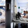
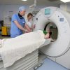
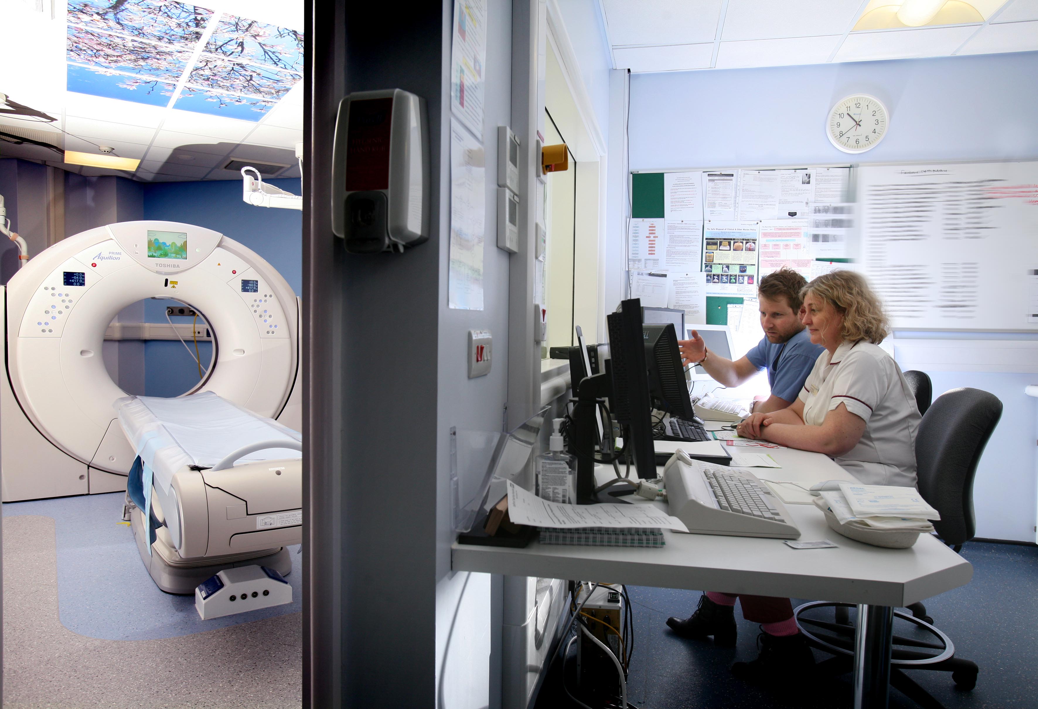
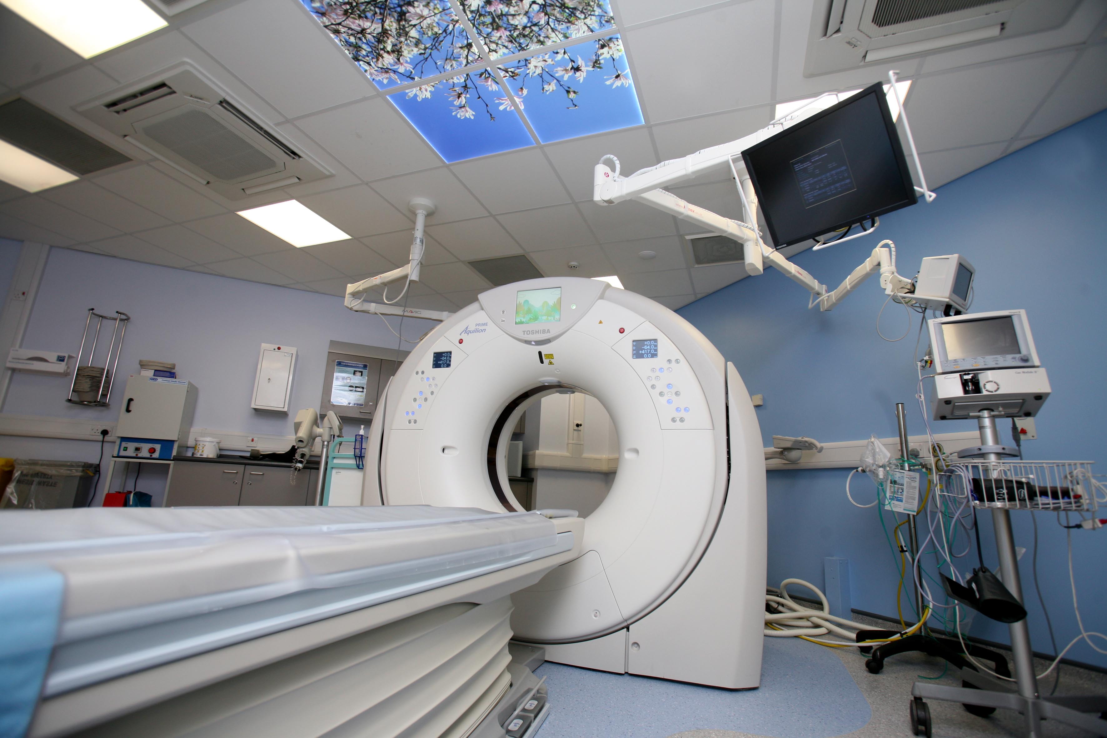
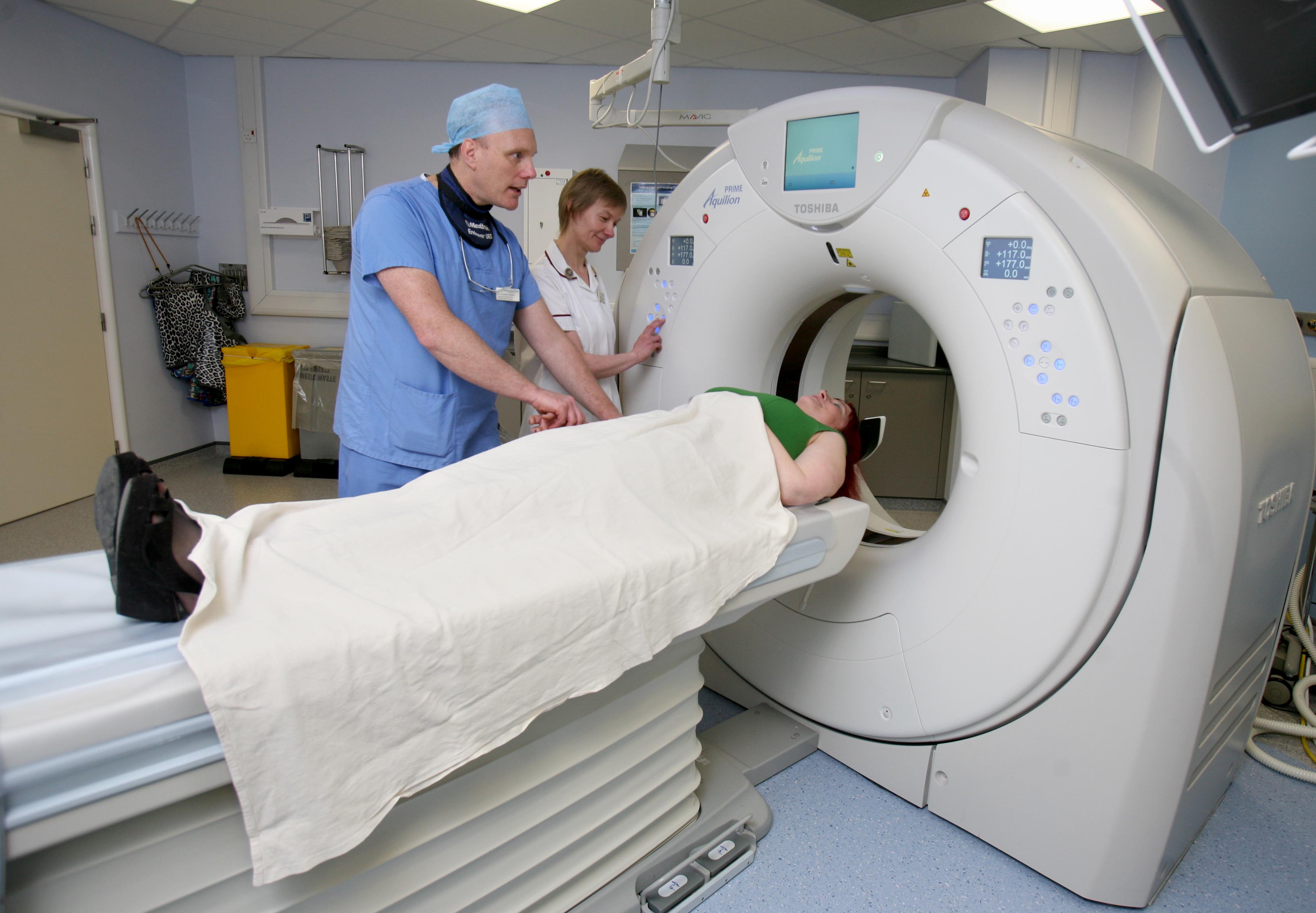
CT Imaging
CT or CAT scan imaging uses x-rays to generate cross-sectional images or ‘slices’ of the body. Patients lie on the CT table which moves through a hole in the scanner. The scan often takes only a few seconds although preparations may take several minutes.
A needle may be placed in your arm to allow injection of contrast or ‘dye’ into a vein. This improves the quality of images and helps the radiologist see more internal details of the body organs. We try to avoid contrast injection if you have poor kidney function, so please let us know if you have any history of kidney failure.
Radiation in very large doses can be harmful, but in general the amount of radiation used in diagnostic imaging is very small. We try to minimise your exposure to radiation by selecting the most appropriate test and technique. We take special care of young patients and pregnant women.
Contrast is usually very safe, but some people may develop a mild skin reaction with itchy lumps called hives or urticaria. This usually improves by itself over 15 to 20 minutes without treatment. Very rarely, patients may have an allergic reaction, like a bad reaction to a bee sting, but the department and staff are equipped to treat these reactions.
Service: Bradford Teaching Hospitals is the reference site for Toshiba’s Aquilion Prime CT scanner and this collaboration with industry has led to the development of low radiation dose and low contrast volume protocols to better patient care.
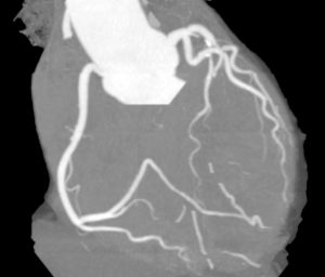
The scanner at St Luke’s is a wide area detector scanner pioneered by Toshiba over 10 years ago, which allows imaging of the heart in just one heartbeart.
The latest addition to the department is the newest 320 detector scanner from Canon, which was installed in March 2018 and brings the latest in scanning technology to Bradford.
Outpatient scanning: Bradford Royal Infirmary: Monday to Friday, 9am-5pm. Many unwell inpatients at BRI are also scanned here and you may experience delays as we try to fit in these patients. Also, urgent patients from A&E may interrupt the workflow through the department but we will try to keep you informed.
St Luke’s Hospital: Outpatients are scanned at St Luke’s Monday to Friday from 8am to 6pm by appointment.
MRI
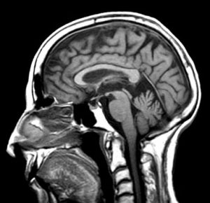
Magnetic Resonance Imaging (MRI) uses a strong magnet and radiowaves to generate images of the body without using x-rays. It is most useful for examining the soft tissues of the brain and spine, areas that are less well visualised with CT scans. Body organs like the liver and bowel, and the bones and joints are also well seen.
MRI scans take longer to acquire than CT scans, often 30–45 minutes for each patient, so working to appointment times is much more important to maximise efficiency. Patients lie on the MRI table inside the scanner for the duration of the scan and this can make patients feel uncomfortable if they have a fear of confirmed spaces. You may visit the department before your scan to avoid having to cancel the scan on the day and to plan any other scans instead.
Service: There are two MR scanners at Bradford Royal Infirmary from Siemens and GE. Both operate at 1.5T strength and permit a wide range of general MR scanning. The movable 1.5T Toshiba scanner at St Luke’s once supported the Olympics in London and has been located on a semi-permanent basis.
Time: Bradford Royal Infirmary – routine scanning takes place between 8am and 8pm. Urgent patients may have to be scanned during the day but we will try to minimise any delays to your scan.
St Luke’s Hospital – routine scanning by appointment between 8am and 8pm.
Fluroscopy and Vascular Imaging
Fluoroscopic screening uses ‘live’ x-rays in real time to see contrast or ‘dye’ moving in the body. This includes watching the action of swallowing, or coating the stomach and small bowel with contrast so we can see the surface.
Live fluoroscopy also allows the radiologist to place needles and injections in joints to aid diagnosis and treat joint pain.
Specialist radiologists and advance practitioners use live angiography to visualise diseased blood vessels in the body and legs by injecting contrast or ‘dye’ through them. Tubes and wires and tiny balloons can be passed through the blood vessels to open up blockages, or metal implants can be inserted under fluoroscopic guidance.
Service: Fluoroscopy is provided at Bradford Royal Infirmary for inpatients and outpatients during normal working hours. Vascular imaging may be performed as a day case requiring a short admission onto a ward.
Nuclear Medicine
Nuclear medicine imaging requires the injection of a small amount of radioactive material into a vein. The specially selected material finds its way to the body tissue we wish to visualise and a special detector camera is used to record the radiation in the organ to generate an image.
Some radioactive materials may be generated on site when needed but many have to be brought in by special order from centres in Manchester and elsewhere for your examination. The dose of radiation we inject is very small and the radioactivity only lasts a short time. It is important to keep appointment times as any unused material is wasted and has to be discarded.
Service: Most scans are performed on an outpatient basis at Bradford Royal Infirmary between 8am and 5pm, Monday to Friday. Expect to be in the Nuclear Medicine Department for two hours or more.


 Head and neck imaging covers disease in the brain, throat, thyroid and salivary glands and sometimes the eyes. CT, MRI and ultrasound are used extensively and many diseases of the neck are available to ultrasound imaging and intervention, including biopsy by Fine Needle Aspiration (FNA) or core biopsy.

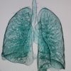
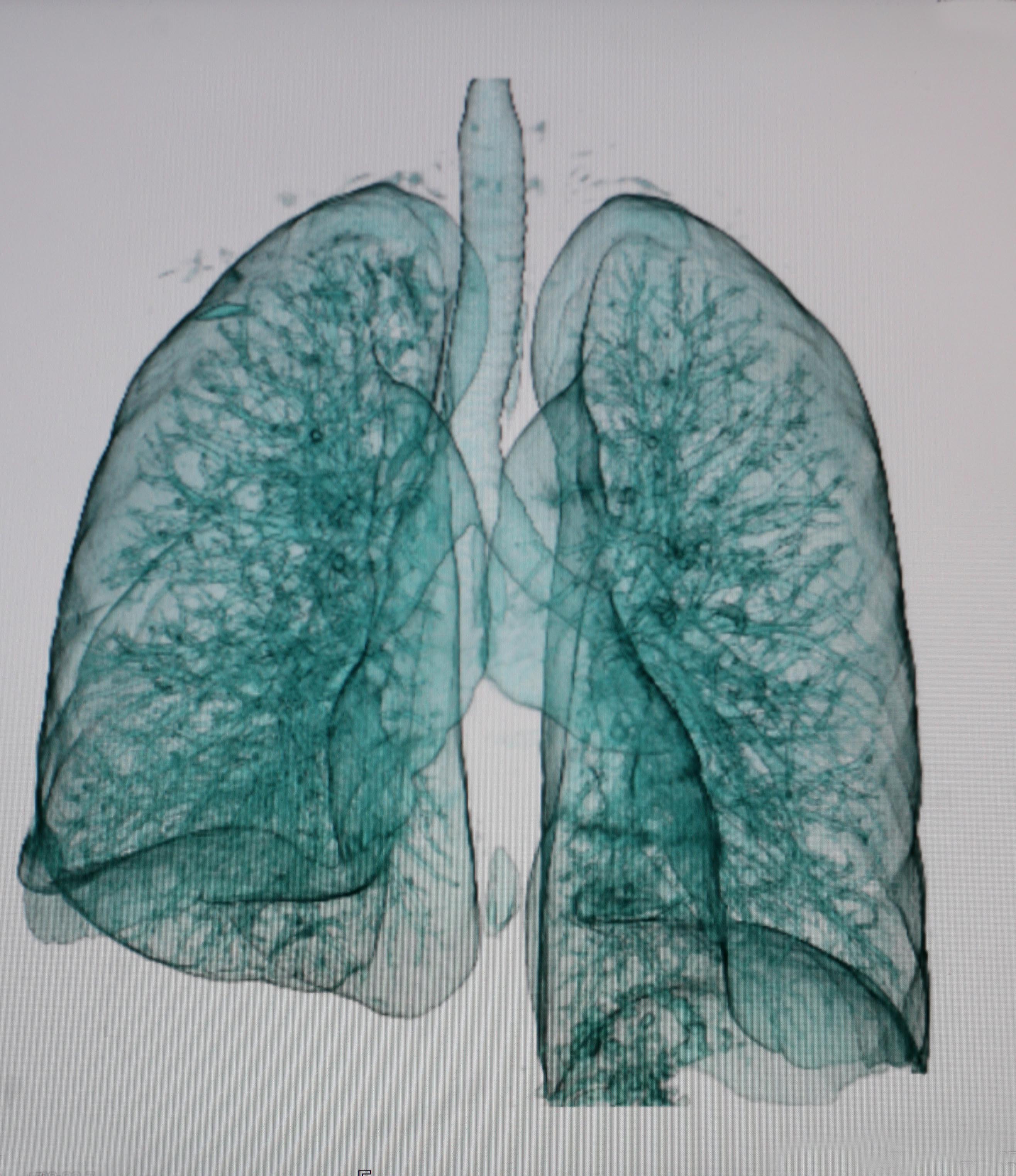
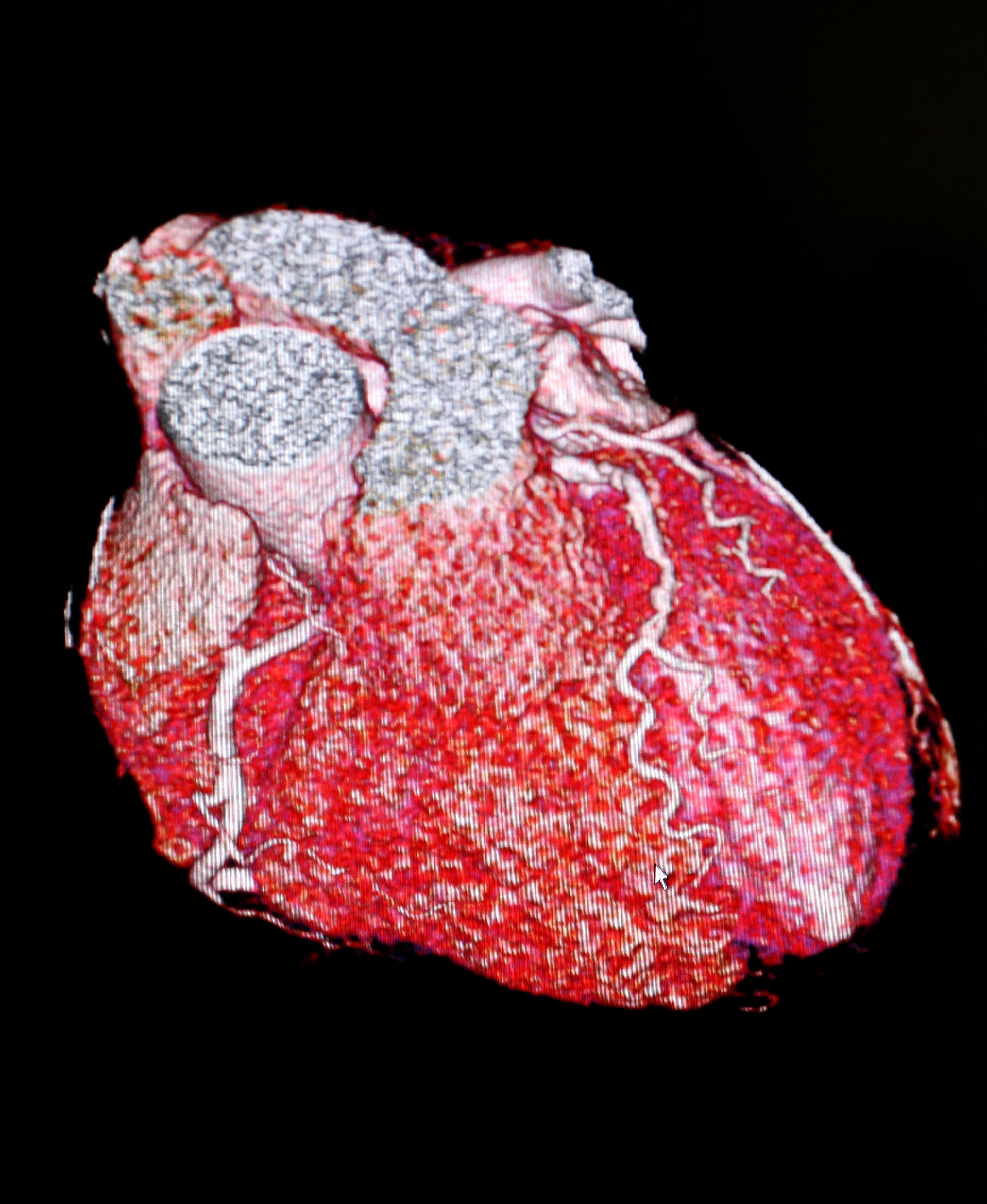
 Thoracic imaging involves visualising the soft tissues of the chest and the lungs in patients with respiratory or chest wall symptoms, often with a chest x-ray in the first instance and then usually by CT imaging.
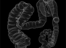 This large specialty covers diseases of the oesophagus stomach, small intestine, large bowel, liver, gallbladder and pancreas. All imaging types are used by all the specialists, in particular MRI for liver pathology, CT colonoscopy for minimally invasive visualisation of the large bowel and MRI of the small bowel.

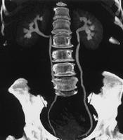 Urogenital imaging mainly covers the kidneys, ureters, bladder and prostate imaging. Kidney stones and tumours are visualised with CT and interventional procedures performed with CT and fluroscopy. Prostate imaging and biopsy with US imaging has increased rapidly.
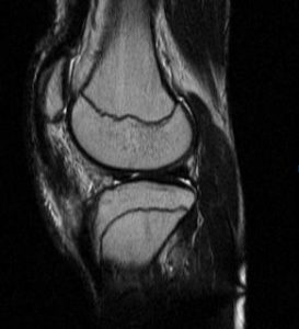 Musculoskeletal imaging of the bones and joints uses plain film radiography initially and subsequently with CT or MR imaging. The spine, especially the lower back, and knee are the most common MR examinations in MSK. Infections or rare bone or muscle tumours are suitably imaged with MR. The joints in the feet and hands and the shoulders are all suited to imaging with Ultrasound, and injections for pain control can also be performed.


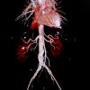
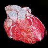
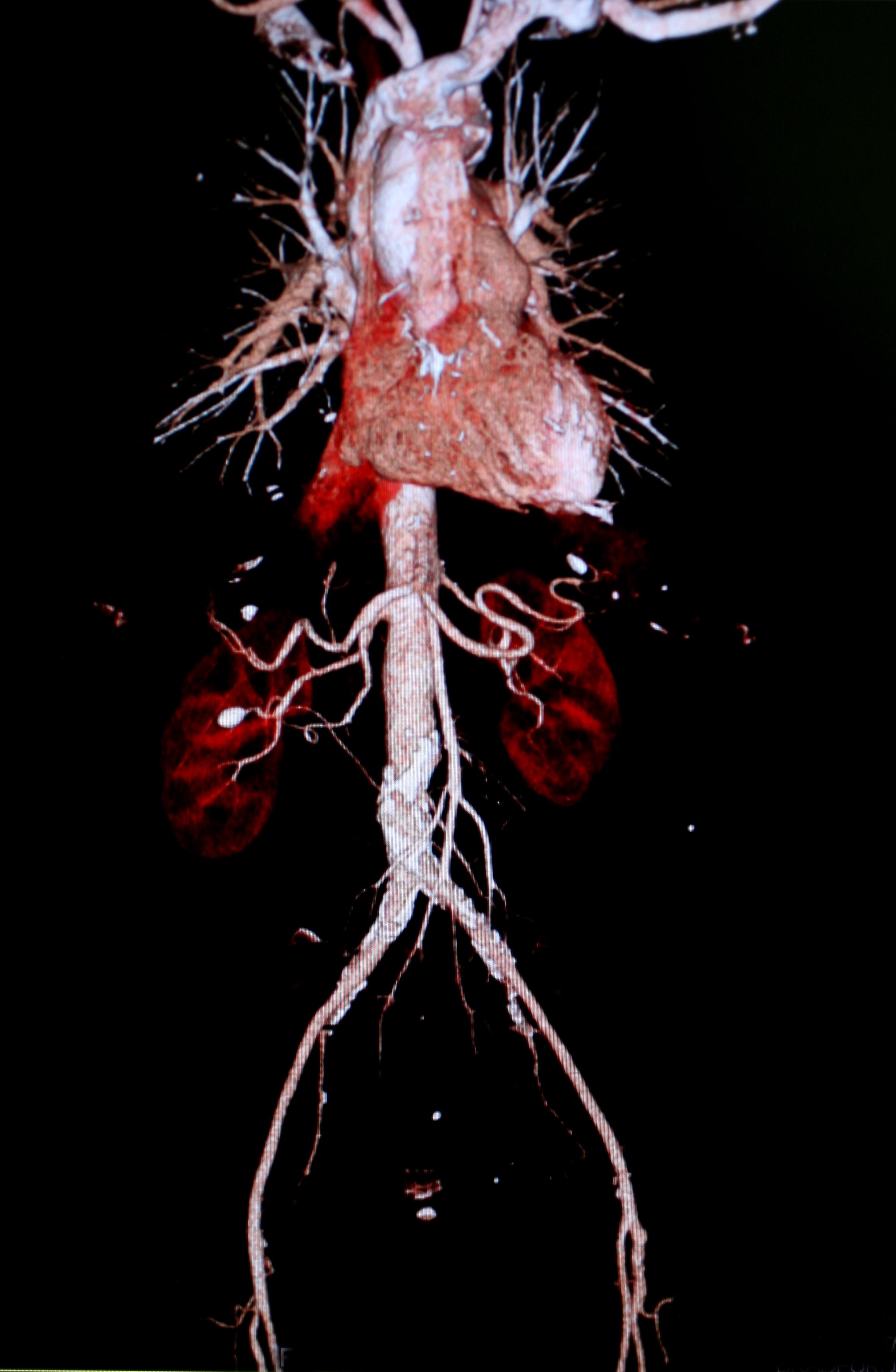
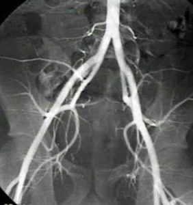 Imaging of the arteries and veins in the body and limbs involves gaining needle access to a blood vessels often at the groin or sometime at the neck, wrist or elbow. Fine wires and tubes are used to inject contrast or dye which can the outline disease in the arteries or veins (angiography). Procedures such as balloon angioplasty or insertion of stents can be performed through these small access points (angioplasty).
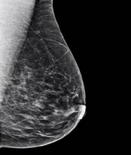 Breast Imaging is provided at the Pennine Breast Services which encompasses Pennine Breast Screening and the Symptomatic Breast Imaging Service.


 Children and young adults are sensitive to radiation so our specialists think carefully before exposing them to x-ray or CT scans.
